
Mount Elizabeth Novena Specialist Centre
38 Irrawaddy Road, #07-41; Singapore 329563
Tel: +65 6254 6678
Fax: +65 6254 6676

Understanding Echocardiograms
Echocardiogram
Ultrasound scans of the heart are called Echocardiograms (frequently shortened to Echo). Because they use high frequency sound waves (ultrasound) to generate the images, they are not harmful and repeat studies can be performed safely.
A doctor may ask for an Echo to be performed for a patient with an abnormal heart trace; chest pain; breathlessness, family history of sudden death and abnormal physical examination.
Echo studies are used to assess
1. the heart function and provides estimates of the internal pressures in the heart
2. the heart structure for enlargement, shunts and abnormal masses.
3. the blood supply to the heart (indirectly) when combined with a stress test
Figure 1 shows an Echo of a heart. which is pumping normally with normal valve function. Figure 2 shows a heart valve which is leaking. The movement of blood is detected using Doppler techniques and is automatically given a colour code by the computer and displayed on the screen. This study shows that there is blood flowing backwards into the left atrium which is the chamber below. The patient will require an operation to repair the leaking valve as the heart will weaken over time due to the additional work it is performing. Figure 3 shows a heart with a large "pericardial effusion", this a collection of fluid around the heart and in this case, the ability of the heart to pump is impaired and a procedure to remove the fluid is indicated. The cause in this patient was a viral infection of the lungs which has also affected the heart function.
Stress Echocardiogram


Figure 5
When combined with a treadmill test, the echocardiogram is able to detect areas of the heart which is not receiving adequate blood flow. Figure 4a and 4b shows the heart function before and after a treadmill test. The patient complained of chest pain soon after the treadmill started and the scan shows that a large section (~ 45%) of the heart was pumping poorly compared to the before scan; which was performed at rest.
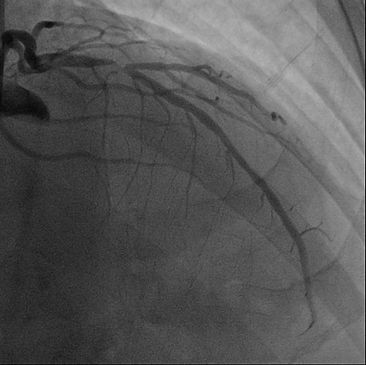
The patient subsequently agreed to undergo a heart angiogram which showed a severe blockage in the Left Anterior Descending (LAD) artery (Figure 4c). The LAD artery would normally supply blood and oxygen to ~ 60% of the heart. Intervention to open up the blockage with a balloon followed by a stent is indicated in this case because a large segment of the heart is at risk everytime the patient exerts himself or is under stress. Figure 4d shows the artery which has been opened up.

Figure 1 / 2 / 3

Figure 4c
If the amount of heart muscle at risk is less than 10%; opening up the blockage is not useful to the patient and in fact may confer more harm. (Figure 5). This is because the procedure itself confers its own risks. In addition to the risk of complications from the procedure, even with modern drug coated stents and new generation of blood thinners (Prasugrel and Ticagrelor), the stent may fail due to a clot forming within the stent (essentially another heart attack) at a rate of about 0.5% per annum.
This may seem counter intuitive but stents and bypasses do not prevent future heart attacks but they do prevent death in the event that one occurs if performed for the appropriate reasons. The main factors in prevention for patients with established coronary heart disease are diet, exercise, cholesterol lowering drugs, stopping smoking, blood thinners and certain blood pressure medications.
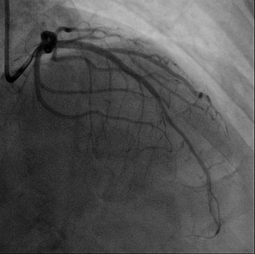
Figure 4d
Figure 4a and 4b

Figure 6a
Figure 6A

Figure 6b
Figure 1
Figure 2
Figure 3
Figure 4C
Figure 4D
A Stress Echocardiogram can also detect problems which are not caused by inadequate blood supply to the heart. In this patient (Figure 6a and 6b); the scan showed that patient had Systolic Anterior Motion (SAM) of the Mitral Valve. Every time the Mitral Valve closes, the valve flips backwards and blocks off the blood that is being pumped out of the heart. There is also significant leakage of blood backwards as well.
The blockage and leakage of blood gets worse when the heart beats faster. Coincidentally, the patient also suffers from asthma and when the patient takes medication for asthma, the symptoms actually worsens due to an increase in the heart rate. Treatment in this case entails taking medication to slow down the heart and in severe cases, open heart surgery to repair the valve.
Stress Echocardiograms are useful as it does not require X-rays or other forms of radiation, it provides information about the heart structure and function; and in particular the heart valves. In addition, it quantifies the amount of heart muscle at risk to aid in the decision to proceed with surgery.
It's downside is that it may be difficult to perform in patients with prior surgery to the chest (e.g. breast implants); lung disease; abnormal bone structure (e,g, Pectus, Scoliosis). However, every patient is unique and most of the time we are able to obtain some useful information from the scan. The ability to pick up significant blockages (called Sensitivity in mathematical term) is excellent but not perfect and most studies will give a Sensitivity of ~ 95% to detect significant blockages of the heart arteries.